45 images of compound microscope with labels
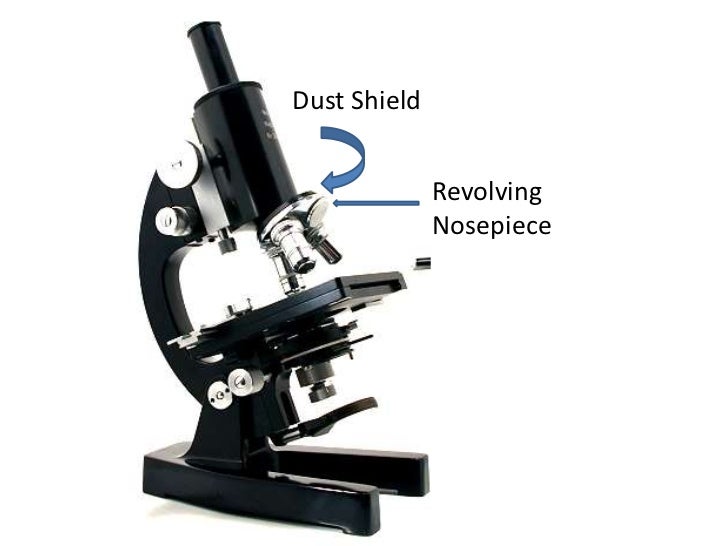
Parts of Stereo Microscope (Dissecting microscope ... Compared to a compound microscope where the objectives attached to the nosepiece can be seen and identified individually (based on color bands and their respective labels), the objectives of a dissecting microscope are located in a cylindrical cone and, therefore, are not directly seen. For the stereo microscope that comes with multiple objective lens sets (fixed power style), the … Ketoconazole Cream vs. Clotrimazole Cream: Dosage, Side Effects The term "ringworm" refers to a fungal infection on the surface of the skin. A physical examination of the affected skin, evaluation of skin scrapings under the microscope, and culture tests can help doctors make the appropriate distinctions. A proper diagnosis is essential to successful treatment.
(PDF) Introduction to Microscopy - ResearchGate • In compound microscope it will be i.e 10 X, f= 16 mm; 40 X, f= 4 mm; 100 X, f= 1.8 mm. • Image produced by objective lens falls on the eyepiece lens serve as objec t. • …

Images of compound microscope with labels
Compound Microscope Parts - Rs' Science Basically, compound microscopes generate magnified images through an aligned pair of the objective lens and the ocular lens. In contrast, "simple microscopes" have only one convex lens and function more like glass magnifiers. [In this figure] Two "antique" microscopes played significant roles in the history of biology. compound microscope parts (labeling) Flashcards - Quizlet Start studying compound microscope parts (labeling). Learn vocabulary, terms, and more with flashcards, games, and other study tools. Parts of a microscope with functions and labeled diagram Parts of a microscope with functions and labeled diagram April 20, 2022 April 19, 2022 by Faith Mokobi Having been constructed in the 16th Century, Microscopes have revolutionalized science with their ability to magnify small objects such as microbial cells, producing images with definitive structures that are identifiable and characterizable.
Images of compound microscope with labels. Multiphoton Microscopy | Nikon’s MicroscopyU The images presented in Figure 7 (a shark choroid plexus stained with fluorescein) provide a comparison of confocal and two-photon microscopy imaging quality. These images were collected at 80-micrometers below the specimen surface, which is the maximal depth allowing sufficient image contrast from this specimen utilizing confocal microscopy ... Cell Types Gizmo Worksheet - Name: Date: Student ... Select the MICROSCOPE tab. Introduction: Complex organisms are made up of smaller units, called cells. Most cells are too small to be seen by the naked eye. Microscopes are used to magnify small objects, so here you will use a compound light microscope to observe the cells of different organisms. Compound Microscope stock vector. Illustration of research ... Illustration about Clearly labeled vector of modern compound microscope. EPS 8 with no gradients or effects, layers labeled for easy editing. Illustration of research, pathologist, magnify - 23417211 Microscope Images Labeled - ncccval Microscope Images Labeled | Virtual Anatomy Lab VAL
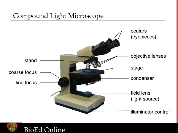
Compound Microscope - BYJUS A microscope with a high resolution and uses two sets of lenses providing a 2-dimensional image of the sample. The term compound refers to the usage of more than one lens in the microscope. Also, the compound microscope is one of the types of optical microscopes. The other type of optical microscope is a simple microscope. PDF Label compound microscope worksheet - Weebly Label compound microscope worksheet ... Light from the illuminator passes through the opening, through the slide and through the lens lens, where the image of the sample is enlarged. Then the enlarged image continues through the body tube microscope to the eyepiece, which further increases the image the viewer then sees. ... Solved Label the image of a compound light microscope ... Experts are tested by Chegg as specialists in their subject area. We review their content and use your feedback to keep the quality high. Transcribed image text: Label the image of a compound light microscope using the terms provided. Compound Microscope Parts, Functions, and Labeled Diagram Compound Microscope Definitions for Labels. Eyepiece (ocular lens) with or without Pointer: The part that is looked through at the top of the compound microscope. Eyepieces typically have a magnification between 5x & 30x. Monocular or Binocular Head: Structural support that holds & connects the eyepieces to the objective lenses.
Microscope Labeling Game - PurposeGames.com About this Quiz. This is an online quiz called Microscope Labeling Game. There is a printable worksheet available for download here so you can take the quiz with pen and paper. This quiz has tags. Click on the tags below to find other quizzes on the same subject. Science. Compound Microscope Labeled Diagram - Quizlet QUESTION. The total magnification of a specimen being viewed with a 10X ocular lens and a 40X objective lens is. 15 answers. QUESTION. a mosquito beats its wings up and down 600 times per second, which you hear as a very annoying 600 Hz sound. if the air outside is 20 C, how far would a sound wave travel between wing beats. 2 answers. Compound Microscope - Diagram (Parts labelled), Principle ... See: Labeled Diagram showing differences between compound and simple microscope parts Structural Components The three structural components include 1. Head This is the upper part of the microscope that houses the optical parts 2. Arm This part connects the head with the base and provides stability to the microscope. Parts of a Compound Microscope - Labeled (with diagrams) A compound microscope is known as a high-power microscope that enables you to achieve a high level of magnification. Smaller specimens can be thoroughly viewed using a compound microscope. Let us take a look at the different parts of a compound microscope and understand each key component.

Robert Hooke - Biography, Facts and Pictures
A&P Flashcards - Quizlet Classify the body location/structure with the correct type of connective tissue by clicking and dragging the labels from column A to the appropriate location in column B. adipose -subcutaneous layer -yellow bone marrow elastic connective-vocal cords-larger artery walls compact bone -bone shafts-spongy bone-ends of long bones-reticular connective -red bone …

Microscope World Blog: Penicillium Under the Microscope
Microscope Types (with labeled diagrams) and Functions A compound microscope: Is used to view samples that are not visible to the naked eye Uses two types of lenses - Objective and ocular lenses Has a higher level of magnification - Typically up to 2000x Is used in hospitals and forensic labs by scientists, biologists and researchers to study micro organisms Compound microscope labeled diagram
Light Microscopy: Instrumentation and Principles | BioEd Online
Recent trends and future of pharmaceutical packaging ... The labels are very similar to destructible labels as mentioned earlier. In this case, the substrate used is of very weak strength paper of low grammage. The paper is also heavily loaded with fillers creating a weak and brittle paper. Labels made from such papers fragment into pieces when attempted to be removed. However, converting it is a very tricky issue when using these …

Microscope World Blog: Lactobacillus under the Microscope
Solved Label the image of a compound light microscope ... Label the image of a compound light microscope using the terms provided. Iris diaphragm lever Eyepiece Light switch Objective lenses Fine adjustment knob Rotating noseplece Stage Slide holder finger Substage illuminator (amp) Mechanical stage control knob One More Course adjustment knob Reset Condenser
Microscopy
Hot and Cold Packs: A Thermochemistry Activity - Carolina.com Compound Microscopes. Popular corded compound microscopes and cordless microscopes for elementary to advanced use. We have the compound microscope you are looking for! Digital Microscopes. Digital microscopes are great for large classroom computer combined instruction. Students can take images, videos, and more. Stereomicroscopes. Stereomicroscopes show …
Parts and functions of a compound microscope
Labelled Diagram of Compound Microscope - Biology Discussion The below mentioned article provides a labelled diagram of compound microscope. Part # 1. The Stand: The stand is made up of a heavy foot which carries a curved inclinable limb or arm bearing the body tube. The foot is generally horse shoe-shaped structure (Fig. 2) which rests on table top or any other surface on which the microscope in kept.

Clipart - microscope with labels
(b) Why both objective and eyepiece of a compound ... Click here👆to get an answer to your question ️ (a) Draw the labelled ray diagram for the formation of image by a compound microscope. Derive an expression for its total magnification (or magnifying power), when the final image is formed at the near point.(b) Why both objective and eyepiece of a compound microscope must have short focal lengths?Draw a ray diagram showing the image ...

Bio World: STRATIFIED EPITHELIUM (COMPOUND EPITHELIUM)
Microscope Drawing And Label - Painting Valley Tags: microscope, label All rights to paintings and other images found on PaintingValley.com are owned by their respective owners (authors, artists), and the Administration of the website doesn't bear responsibility for their use.
Post a Comment for "45 images of compound microscope with labels"