41 diagram of a human cell with labels
Male Reproductive System: Labeled Diagram of Organs ... The female gamete is the ovum, and is the largest human cell that is ovulated in the female reproductive system. The male reproductive system is composed of both external and internal organs that ... Animal Cell Diagram with Label and Explanation: Cell ... Below is the diagram of the animal cell which shows the organelles present in it. The cell is covered with cytoplasm which consists of cell organelles in it. The nucleus is covered with a rough Endoplasmic Reticulum and other organelles each designed for a specific purpose.
Cell: Structure and Functions (With Diagram) Eukaryotic Cells: 1. Eukaryotes are sophisticated cells with a well defined nucleus and cell organelles. 2. The cells are comparatively larger in size (10-100 μm). 3. Unicellular to multicellular in nature and evolved ~1 billion years ago. 4. The cell membrane is semipermeable and flexible. 5. These cells reproduce both asexually and sexually.

Diagram of a human cell with labels
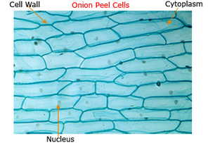
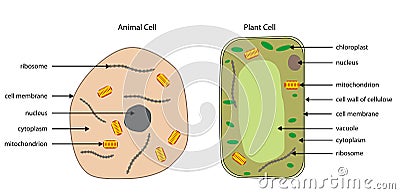
Labeled Plant Cell With Diagrams - Science Trends Plant cells contain many organelles such as ribosomes, the nucleus, the plasma membrane, the cell wall, mitochondria, and chloroplasts. In addition, plant cells differ from animal cells in a number of key ways. Examining a diagram of the plant cell will help make the differences clearer. Let's go over the individual components of plant cells ... Human Cells Printables and Diagrams - The Successful ... These cells include: leukocytes, haematids, thrombocytes, ovum, sperm, sarcomeres, enterocytes, neurons, osteocytes, hepatocytes. They will learn the parts of a cell thanks to a labeled diagram. They will get to see what blood looks like under a microscope without needing to own a microscope. They get to color a cell and then label the parts. Human Cell Diagram, Parts, Pictures, Structure and Functions One of the few cells in the human body that lacks almost all organelles are the red blood cells. The main organelles are as follows : cell membrane endoplasmic reticulum Golgi apparatus lysosomes mitochondria nucleus perioxisomes microfilaments and microtubules Diagram of the human cell illustrating the different parts of the cell. Cell Membrane
Diagram of a human cell with labels. Labeled Diagram of the Human Lungs - Bodytomy Given below is a labeled diagram of the human lungs followed by a brief account of the different parts of the lungs and their functions. Each lung is enclosed inside a sac called pleura, which is a double-membrane structure formed by a smooth membrane called serous membrane. Labeled Diagram of the Human Kidney - Bodytomy Labeled Diagram of the Human Kidney The human kidneys house millions of tiny filtration units called nephrons, which enable our body to retain the vital nutrients, and excrete the unwanted or excess molecules as well as metabolic wastes from the body. In addition, they also play an important role in maintaining the water balance of our body. What Is Going On Inside That Cell? | Human cell diagram ... cell, in biology, the basic membrane-bound unit that contains the fundamental molecules of life and of which all living things are composed. A single cell is often a complete organism in itself, such as a bacterium or yeast. Other cells acquire specialized functions as they mature. Anatomy (Human Body) Labeling - Exploring Nature Muscles of the Leg and Foot Labeling Page. Muscles of the Neck, Chest and Thorax Labeling Page. Muscles of the Neck, Shoulders and Thorax (Posterior) Labeling. Muscles of the Posterior Body Labeling (HS-Adult) Muscles of the Thigh and Hip (Anterior) Labeling. Muscles of the Thigh and Hip (Posterior) Labeling. Nerve Cell (Neuron) Labeling Page.
Circulatory System Labeled Diagram stock illustrations Browse 154 circulatory system labeled diagram stock illustrations and vector graphics available royalty-free, or start a new search to explore more great stock images and vector art. Newest results. Heart Poster Heart anatomical poster containing detailed information about the human heart. Cell Model Label the Parts Diagram | Quizlet Start studying Cell Model Label the Parts. Learn vocabulary, terms, and more with flashcards, games, and other study tools. Pin by Lahu deore on Dc. | Human cell diagram, Cell ... This diagram of a human skeleton is labeled with 12 major bones, from skull to fibula. D Nicole Science Tissue Biology Anatomy And Physiology Textbook Histology Slides Biology College Basic Physics Chemistry Lessons Nursing School Notes 6th Grade Science Medical Coding 18" by 24" Laminated Wall Chart A My Airtel Nursing Diagram of human skin structure - Science Learning Hub Diagram of human skin structure. Add to collection. + Create new collection. Tweet. Rights: University of Waikato Published 1 February 2011 Size: 100 KB Referencing Hub media. The epidermis is a tough coating formed from overlapping layers of dead skin cells.
A Well-labelled Diagram Of Animal Cell With Explanation Well-Labelled Diagram of Animal Cell The Cell Organelles are membrane-bound, present within the cells. There are various organelles present within the cell and are classified into three categories based on the presence or absence of membrane. Listed below are the Cell Organelles of an animal cell along with their functions. Learn the parts of a cell with diagrams and cell quizzes For this exercise we'll start with an image of a cell diagram ready labeled. Study this and make sure that you're clear about which structure is found where. Cell diagram unlabeled It's time to label the cell yourself! As you fill in the cell structure worksheet, remember the functions of each part of the cell that you learned in the video. Cell Membrane Diagram Labeled : Functions and Diagram Cell Membrane Diagram Labeled. Monday, March 22nd 2021. | Diagram. Cell Membrane Diagram. There are no organelles in the prokaryotic cells, i.e., they have no internal membrane systems. While lipids help to give membranes their flexibility, proteins monitor and maintain. We all keep in mind that the human body is very elaborate and a technique ... Draw Structure of human cell and label? - Answers The purpose of the cell wall in a plant cell is to give the cell structure and shape. Animal and human cells have no specific shape or structure. People also asked
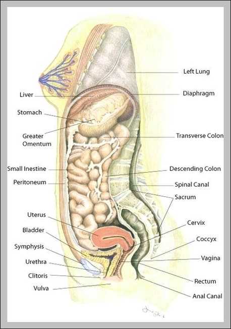
uterus location | Anatomy System - Human Body Anatomy diagram and chart images
Animal Cells: Labelled Diagram, Definitions, and Structure Animal Cells Organelles and Functions. A double layer that supports and protects the cell. Allows materials in and out. The control center of the cell. Nucleus contains majority of cell's the DNA. Popularly known as the "Powerhouse". Breaks down food to produce energy in the form of ATP.

Xtra! Xtra! Read All About It Newspaper!: Biology: Learning About Cells
Human Cell Organelles Labeling Diagram - Quizlet Human Cell Organelles Labeling STUDY Learn Flashcards Write Spell Test PLAY Match Gravity Created by Mackenna_Rios5 Terms in this set (8) Vesicles Transports molecules between organelles and the cell membrane Ribosome Makes Protein Mitochondria Makes ATP Smooth ER Makes lipids and vesicles Lysosomes
PRACTICAL BOOKLET - BIOLOGY4ISC
Blood Cell Diagram Stock Photos, Pictures & Royalty-Free ... Bone marrow Blood stem cell is an immature cell that can develop into all types of blood cells, including white blood cells, red blood cells, and platelets. Blood stem cells are found in the peripheral blood and the bone marrow. Also called hematopoietic stem cell. 3d render blood cell diagram stock pictures, royalty-free photos & images.

Luke's Place | This blog is about my school year and myself.
Skeletal System - Labeled Diagrams of the Human Skeleton The skeletal system's cell matrix acts as our calcium bank by storing and releasing calcium ions into the blood as needed. Proper levels of calcium ions in the blood are essential to the proper function of the nervous and muscular systems. Bone cells also release osteocalcin, a hormone that helps regulate blood sugar and fat deposition.
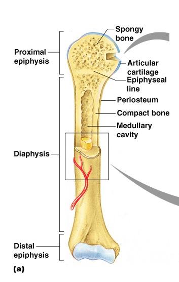
Print Exercise 9: Overview of the Skeleton: Classification and Structure of Bones and Cartilages ...
Heart Diagram with Labels and Detailed Explanation - BYJUS Diagram of Heart. The human heart is the most crucial organ of the human body. It pumps blood from the heart to different parts of the body and back to the heart. The most common heart attack symptoms or warning signs are chest pain, breathlessness, nausea, sweating etc. The diagram of heart is beneficial for Class 10 and 12 and is frequently ...

Follow me: Arts
Label Diagram Human Body Stock Illustrations - Dreamstime Download 161 Label Diagram Human Body Stock Illustrations, Vectors & Clipart for FREE or amazingly low rates! New users enjoy 60% OFF. 185,925,055 stock photos online.

Science
Human Heart Diagram Labeled | Science Trends The human heart is an organ responsible for pumping blood through the body, moving the blood (which carries valuable oxygen) to all the tissues in the body. Without the heart, the tissues couldn't get the oxygen they need and would die. Along with lymphatic vessels, the blood, blood vessels, and lymph, the heart composes the circulatory system of the body.

Human Cell Diagram To Label - General Wiring Diagram
A Labeled Diagram of the Animal Cell and its Organelles A Labeled Diagram of the Animal Cell and its Organelles. There are two types of cells - Prokaryotic and Eucaryotic. Eukaryotic cells are larger, more complex, and have evolved more recently than prokaryotes. Where, prokaryotes are just bacteria and archaea, eukaryotes are literally everything else. From amoebae to earthworms to mushrooms, grass ...

Anatomy of a human cell with labels for individual organelles | Organelles, Medical illustration ...
PDF Human Cell Diagram, Parts, Pictures, Structure and Functions Diagram of the human cell illustrating the different parts of the cell. Cell Membrane The cell membraneis the outer coating of the cell and contains the cytoplasm, substances within it and the organelle. It is a double-layered membrane composed of proteins and lipids.

Animal Cell Diagram. Stock Illustration - Image: 68054896
Human Cell Diagram, Parts, Pictures, Structure and Functions One of the few cells in the human body that lacks almost all organelles are the red blood cells. The main organelles are as follows : cell membrane endoplasmic reticulum Golgi apparatus lysosomes mitochondria nucleus perioxisomes microfilaments and microtubules Diagram of the human cell illustrating the different parts of the cell. Cell Membrane

a. Cell : unit of function - BIOLOGY4ISC
Human Cells Printables and Diagrams - The Successful ... These cells include: leukocytes, haematids, thrombocytes, ovum, sperm, sarcomeres, enterocytes, neurons, osteocytes, hepatocytes. They will learn the parts of a cell thanks to a labeled diagram. They will get to see what blood looks like under a microscope without needing to own a microscope. They get to color a cell and then label the parts.

cell labeled | HUMAN ANATOMY & PHYSIOLOGY | Pinterest
Labeled Plant Cell With Diagrams - Science Trends Plant cells contain many organelles such as ribosomes, the nucleus, the plasma membrane, the cell wall, mitochondria, and chloroplasts. In addition, plant cells differ from animal cells in a number of key ways. Examining a diagram of the plant cell will help make the differences clearer. Let's go over the individual components of plant cells ...

Animal Cell Coloring Key | Animal cells worksheet, Color worksheets, Animal cell structure

Brain Diagram - Labeled - Color - Tim's Printables
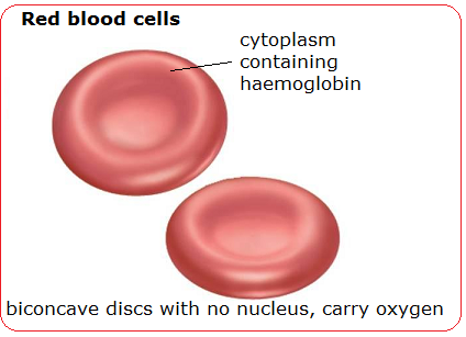
Blood cells - structure and functions - Biology Notes for IGCSE 2014
Diagrams Of Animal And Plant Cells Stock Vector - Image: 45385330
Post a Comment for "41 diagram of a human cell with labels"